RECENT PROJECTS
Machine learning methods for Medical Image Analysis
We developed a cost efficient framework for image segmentation that dramatically reduces the cost of generating labeled training data by intelligently selecting few samples to be labeled by achieving similar performance as to labeling the entire data set.
Sourati, J., Gholipour, A., Dy, J.G., Tomas-Fernandez, X., Kurugol, S. and Warfield, S.K., 2019.
Intelligent labeling based on fisher information for medical image segmentation using deep learning.
IEEE transactions on medical imaging,
38(11), pp.2642-2653.
We developed a deep learning technique that integartes a shape prior into a CNN for image segmentation. This methods improves performance when weak labels are used for segmentation.
Mortazi, A., Khosravan, N., Torigian, D.A., Kurugol, S. and Bagci, U., 2019, October.
Weakly Supervised Segmentation by a Deep Geodesic Prior. In
International Workshop on Machine Learning in Medical Imaging (pp. 238-246). Springer, Cham.
Simultaneous Motion and Distortion Corrected Diffusion-weighted MRI of Kidneys
Motion and distortion are reducing the robustness of quantitative diffusion-weighted MRI in kidneys. Motion correction methods such as respiratory gating dramatically increase the imaging time. Also, currently available distortion correction methods cannot fully correct for distortion in the presence of motion because distortion field changes with motion.
We have recently introduced a motion and distortion corrected DW-MRI method for brain and kidneys that improve the precision of parameters of the IVIM and DTI models.
Related Publications:
Afacan, O., Hoge, W.S., Wallace, T.E., Gholipour, A., Kurugol, S. and Warfield, S.K., 2020.
Simultaneous Motion and Distortion Correction Using Dual‐Echo Diffusion‐Weighted MRI.
Journal of Neuroimaging.
Coll‐Font J, Afacan O, Hoge, T.E., Gholipour, A., Marami, B.,Warfield, S.K., Kurugol, S. 2020. Simultaneous distortion and motion correction in abdominal DW-MRI using dual echo EPI and slice-to-volume registration. Proceedings of Int. Society of Magnetic Resonance in Medicine.
Motion robust DCE-MRI for functional imaging of kidneys without sedation in infants
We have developed a motion-robust DCE-MRI technique to compensate for the bulk motion of baby imaged during sleep in the scanner. Our bulk motion detection technique removes the corrupted volumes, and our motion correction technique realigns the volumes acquired with dynamic radial VIBE sequence. Our methods enables non-sedated imaging of infants to compute their differential renal function and glomerular filtration rate, the imaging markers used for important clinical decisions.
Related Publications:
Kurugol, S., Afacan, O., Lee, R. S., Seager, C. M., Ferguson, M. A., Stein, D. R., ... & Chow, J. S. (2020).
Prospective pediatric study comparing glomerular filtration rate estimates based on motion-robust dynamic contrast-enhanced magnetic resonance imaging and serum creatinine (eGFR) to 99m Tc DTPA.
Pediatric radiology, 1-8.
Kurugol, S., Seager, C.M., Thaker, H., Coll-Font, J., Afacan, O., Nichols, R.C., Warfield, S.K., Lee, R.S. and Chow, J.S., 2019.
Feed and wrap magnetic resonance urography provides anatomic and functional imaging in infants without anesthesia.
Journal of Pediatric Urology.
Diffusion-weighted MRI in abdomen- Intravoxel Incoherent Motion Model (IVIM)
Spatially-Constrained IVIM Model (SCIM) and Spatially Constrained Probabislistic Incohenrent Motion Model (SPIM)
Diffusion weighted MRI signal in abdomen is is often modelled by single exponential model with a decay rate parameter, i.e. ADC. However, ADC parameter does not fully capture the signal measured by DW-MRI and therefore different choices of diffusion weightings (b-values) result in different ADC values. IVIM model is a more accurate model of the signal decay that considers both the slow diffusion due to movement of water molecules and also fast diffusion related to microcapillary perfusion. Unfortunately, IVIM model parameter estimation is not robust due to several factors including motion, low SNR and lack of enough data acquired at low b-values to estimate fast diffusion parameter and the fast diffusion fraction (f) parameter. In order to account for this problem our group developed a series of techniques:
1) Spatially-constratined IVIM (SCIM) model for abdomen: This model achieves improved precision when estimating IVIM model parameters by regularizing over the space when estimating the parameters for each voxel.
2) Probabilistic Model of Slow and Fast Diffusion: The slow and fast diffusion happens at various scales and therefore it is more accurate to model each parameter with a probability distirbution instead of assuming that they have a single constant value. This approach results in more accurate modeling. We use a variable projection method for solving for the parameters which improves the accurqacy and precision of the parameter estimates,
3) Spatially-constratined Probability Model of Incoherent Motion (SPIM): This model improves the robustness and precision of estimated parameters by regularizing over the space when estimating the parameters for each voxel.
4) Correcting for the Effect of Motion using Simultaneous Image Registration and Model Estimation (SIR-ME) in Abdominal DW-MRI: In abdominal DW-MRI, images are often acquired during free breathing in clinical practice because alternative techniques such as respiratory gating increases the scan time. This results in motion due to respiration, motility of bowel results in misalignment between images and reduces the accuracy of parameter estimates. We therefore developed an image regsitration based motion correction that utilizas the dependence of images through the signal decay equation into account when performing the registration for motion correction.
Diffusion-weighted MRI for Crohn's disease
Crohn's Disease: Crohn's disease (CD) is a chronic inflammatory bowel disease, which has relapsing, and remitting clinical course. Long-standing inflammation can result in bowel obstruction, stricture, fistula, abscess and an increased risk for small and large bowel malignancy. Identifying disease stage is crucial for determining the correct treatment options. Magnetic resonance imaging (MRI) allows accurate assessment of Crohn’s disease (CD), but requires gadolinium injection, which poses risks. Diffusion-weighted (DW-MRI) yields comparable performances in small bowel CD without requiring gadolinium injection.
Spatially-Constrained Probability Distribution Model of Incoherent Motion (SPIM) for Abdominal Diffusion-Weighted MRI: Quantitative diffusion-weighted MR imaging (DW-MRI) of the body enables characterization of the tissue microenvironment by measuring variations in the mobility of water molecules. The diffusion signal decay model parameters are increasingly used to evaluate various diseases of abdominal organs such as the liver and spleen. However, previous signal decay models (i.e., mono-exponential, bi-exponential intra-voxel incoherent motion (IVIM) and stretched exponential models) only provide insight into the average of the distribution of the signal decay rather than explicitly describe the entire range of diffusion scales. In this work, we propose a probability distribution model of incoherent motion that uses a mixture of Gamma distributions to fully characterize the multi-scale nature of diffusion within a voxel. Further, we improve the robustness of the distribution parameter estimates by integrating spatial homogeneity prior into the probability distribution model of incoherent motion (SPIM) and by using the fusion bootstrap solver (FBM) to estimate the model parameters. We evaluated the improvement in quantitative DW-MRI analysis achieved with the SPIM model in terms of accuracy, precision and reproducibility of parameter estimation in both simulated data and in 68 abdominal in-vivo DW-MRIs. Our results show that the SPIM model not only substantially reduced parameter estimation errors by up to 26%; it also significantly improved the robustness of the parameter estimates (paired Students t-test, p
Motion Compensated Abdominal Diffusion Weighted MRI by Simultaneous Image Registration and Model Estimation (SIR-ME): Non-invasive characterization of water molecule's mobility variations by quantitative analysis of diffusion-weighted MRI (DW-MRI) signal decay in the abdomen has the potential to serve as a biomarker in gastrointestinal and oncological applications. Accurate and reproducible estimation of the signal decay model parameters is challenging due to the presence of respiratory, cardiac, and peristalsis motion. Independent registration of each b-value image to the b-value=0 s/mm2 image prior to parameter estimation might be sub-optimal because of the low SNR and contrast difference between images of varying b-value. In this work, we introduce a motion-compensated parameter estimation framework that simultaneously solves image registration and model estimation (SIR-ME) problems by utilizing the interdependence of acquired volumes along the diffusion weighting dimension. We evaluated the improvement in model parameters estimation accuracy using 16 in-vivo DW-MRI data sets of Crohn's disease patients by comparing parameter estimates obtained using the SIR-ME model to the parameter estimates obtained by fitting the signal decay model to the acquired DW-MRI images. The proposed SIR-ME model reduced the average root-mean-square error between the observed signal and the fitted model by more than 50%. Moreover, the SIR-ME model estimates discriminate between normal and abnormal bowel loops better than the standard parameter estimates.
PREVIOUS PROJECTS
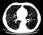
Modeling Spatial Distribution of Emphysema Patterns in Volumetric CTs of COPD Patients:
- Classification of Lung Regions into Patterns of Emphysema Progression Levels
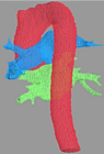
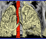
Automated Quantification of Cardiovascular Disease Phenotypes in Volumetric CTs of Chronic Obstructive Pulmonary Disease (COPD) Patients: This project involved development of algorithms for segmentation of large vessels and extraction of morphological and calcification measures as imaging markers of cardiovascular disease. Specifically, I developed an automated aorta segmentation and aortic calcification detection algorithm. I also developed and algorithm to extract aorta morphology measures (e.g. Arch width, radius and cross sectional area, tortuosity, curvature)
3-D/4-D Segmentation of Structures in CT Images for Radiotherapy Planning: This project involved development of segmentation algorithms for radiotherapy planning. Specifically, I developed a knowledge based esophagus segmentation algorithm that uses a novel global-local shape model and a spatial dependency model that I introduced to solve this difficult problem.
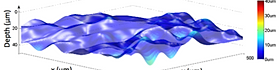
Towards Automated Analysis of Confocal Microscopy Images of Human Skin for Early Cancer Detection: Confocal Microscopy allows early skin cancer detection without the need for biopsy followed by histopathology. In this work, I developed automated image analysis algorithms for confocal microscopy images: 1) A locally smooth SVM classification algorithm for detection of the skin layers; 2) Hybrid Sequential Segmentation and Locally Smooth Classification Approach to Detect Dermal Epidermal Junction.