Publications

E

Coulter, Michael, Cristina Dorobantu, Gerrald Lodewijk, François Delalande, Sarah Cianferani, Vijay Ganesh, Richard Smith, et al. 2018. “The ESCRT-III Protein CHMP1A Mediates Secretion of Sonic Hedgehog on a Distinctive Subtype of Extracellular Vesicles”. Cell Rep 24 (4): 973-986.e8. https://doi.org/10.1016/j.celrep.2018.06.100.
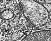
Endosomal sorting complex required for transport (ESCRT) complex proteins regulate biogenesis and release of extracellular vesicles (EVs), which enable cell-to-cell communication in the nervous system essential for development and adult function. We recently showed human loss-of-function (LOF) mutations in ESCRT-III member CHMP1A cause autosomal recessive microcephaly with pontocerebellar hypoplasia, but its mechanism was unclear. Here, we show Chmp1a is required for progenitor proliferation in mouse cortex and cerebellum and progenitor maintenance in human cerebral organoids. In Chmp1a null mice, this defect is associated with impaired sonic hedgehog (Shh) secretion and intraluminal vesicle (ILV) formation in multivesicular bodies (MVBs). Furthermore, we show CHMP1A is important for release of an EV subtype that contains AXL, RAB18, and TMED10 (ART) and SHH. Our findings show CHMP1A loss impairs secretion of SHH on ART-EVs, providing molecular mechanistic insights into the role of ESCRT proteins and EVs in the brain.
Lee, Wei-Chung Allen, and Elly Nedivi. 2002. “Extended Plasticity of Visual Cortex in Dark-Reared Animals May Result from Prolonged Expression of Cpg15-Like Genes.”. J Neurosci 22 (5): 1807-15.
cpg15 is an activity-regulated gene that encodes a membrane-bound ligand that coordinately regulates growth of apposing dendritic and axonal arbors and the maturation of their synapses. These properties make it an attractive candidate for participating in plasticity of the mammalian visual system. Here we compare cpg15 expression during normal development of the rat visual system with that seen in response to dark rearing, monocular blockade of retinal action potentials, or monocular deprivation. Our results show that the onset of cpg15 expression in the visual cortex is coincident with eye opening, and it increases until the peak of the critical period at postnatal day 28 (P28). This early expression is independent of both retinal activity and visual experience. After P28, a component of cpg15 expression in the visual cortex, lateral geniculate nucleus (LGN), and superior colliculus (SC) develops a progressively stronger dependence on retinally driven action potentials. Dark rearing does not affect cpg15 mRNA expression in the LGN and SC at any age, but it does significantly affect its expression in the visual cortex from the peak of the critical period and into adulthood. In dark-reared rats, the peak level of cpg15 expression in the visual cortex at P28 is lower than in controls. Rather than showing the normal decline with maturation, these levels are maintained in dark-reared animals. We suggest that the prolonged plasticity in the visual cortex that is seen in dark-reared animals may result from failure to downregulate genes such as cpg15 that could promote structural remodeling and synaptic maturation.

D

Lee, Wei-Chung Allen, Hayden Huang, Guoping Feng, Joshua Sanes, Emery Brown, Peter So, and Elly Nedivi. (2006) 2006. “Dynamic Remodeling of Dendritic Arbors in GABAergic Interneurons of Adult Visual Cortex.”. PLoS Biol 4 (2): e29. https://doi.org/10.1371/journal.pbio.0040029.
Despite decades of evidence for functional plasticity in the adult brain, the role of structural plasticity in its manifestation remains unclear. To examine the extent of neuronal remodeling that occurs in the brain on a day-to-day basis, we used a multiphoton-based microscopy system for chronic in vivo imaging and reconstruction of entire neurons in the superficial layers of the rodent cerebral cortex. Here we show the first unambiguous evidence (to our knowledge) of dendrite growth and remodeling in adult neurons. Over a period of months, neurons could be seen extending and retracting existing branches, and in rare cases adding new branch tips. Neurons exhibiting dynamic arbor rearrangements were GABA-positive non-pyramidal interneurons, while pyramidal cells remained stable. These results are consistent with the idea that dendritic structural remodeling is a substrate for adult plasticity and they suggest that circuit rearrangement in the adult cortex is restricted by cell type-specific rules.
Lee, Wei-Chung Allen, Jerry Chen, Hayden Huang, Jennifer Leslie, Yael Amitai, Peter So, and Elly Nedivi. 2008. “A Dynamic Zone Defines Interneuron Remodeling in the Adult Neocortex.”. Proc Natl Acad Sci U S A 105 (50): 19968-73. https://doi.org/10.1073/pnas.0810149105.
The contribution of structural remodeling to long-term adult brain plasticity is unclear. Here, we investigate features of GABAergic interneuron dendrite dynamics and extract clues regarding its potential role in cortical function and circuit plasticity. We show that remodeling interneurons are contained within a "dynamic zone" corresponding to a superficial strip of layers 2/3, and remodeling dendrites respect the lower border of this zone. Remodeling occurs primarily at the periphery of dendritic fields with addition and retraction of new branch tips. We further show that dendrite remodeling is not intrinsic to a specific interneuron class. These data suggest that interneuron remodeling is not a feature predetermined by genetic lineage, but rather, it is imposed by cortical laminar circuitry. Our findings are consistent with dynamic GABAergic modulation of feedforward and recurrent connections in response to top-down feedback and suggest a structural component to functional plasticity of supragranular neocortical laminae.

A

Lee, Wei-Chung Allen, Vincent Bonin, Michael Reed, Brett Graham, Greg Hood, Katie Glattfelder, and Clay Reid. 2016. “Anatomy and Function of an Excitatory Network in the Visual Cortex.”. Nature 532 (7599): 370-4. https://doi.org/10.1038/nature17192.
Circuits in the cerebral cortex consist of thousands of neurons connected by millions of synapses. A precise understanding of these local networks requires relating circuit activity with the underlying network structure. For pyramidal cells in superficial mouse visual cortex (V1), a consensus is emerging that neurons with similar visual response properties excite each other, but the anatomical basis of this recurrent synaptic network is unknown. Here we combined physiological imaging and large-scale electron microscopy to study an excitatory network in V1. We found that layer 2/3 neurons organized into subnetworks defined by anatomical connectivity, with more connections within than between groups. More specifically, we found that pyramidal neurons with similar orientation selectivity preferentially formed synapses with each other, despite the fact that axons and dendrites of all orientation selectivities pass near (5 μm) each other with roughly equal probability. Therefore, we predict that mechanisms of functionally specific connectivity take place at the length scale of spines. Neurons with similar orientation tuning formed larger synapses, potentially enhancing the net effect of synaptic specificity. With the ability to study thousands of connections in a single circuit, functional connectomics is proving a powerful method to uncover the organizational logic of cortical networks.