Publications

W

Tobin, William, Rachel Wilson, and Wei-Chung Allen Lee. 2017. “Wiring Variations That Enable and Constrain Neural Computation in a Sensory Microcircuit”. Elife 6. https://doi.org/10.7554/eLife.24838.
Neural network function can be shaped by varying the strength of synaptic connections. One way to achieve this is to vary connection structure. To investigate how structural variation among synaptic connections might affect neural computation, we examined primary afferent connections in the Drosophila olfactory system. We used large-scale serial section electron microscopy to reconstruct all the olfactory receptor neuron (ORN) axons that target a left-right pair of glomeruli, as well as all the projection neurons (PNs) postsynaptic to these ORNs. We found three variations in ORN→PN connectivity. First, we found a systematic co-variation in synapse number and PN dendrite size, suggesting total synaptic conductance is tuned to postsynaptic excitability. Second, we discovered that PNs receive more synapses from ipsilateral than contralateral ORNs, providing a structural basis for odor lateralization behavior. Finally, we found evidence of imprecision in ORN→PN connections that can diminish network performance.
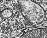
Hildebrand, David Grant Colburn, Marcelo Cicconet, Russel Miguel Torres, Woohyuk Choi, Tran Minh Quan, Jungmin Moon, Arthur Willis Wetzel, et al. 2017. “Whole-Brain Serial-Section Electron Microscopy in Larval Zebrafish”. Nature. https://doi.org/10.1038/nature22356.
High-resolution serial-section electron microscopy (ssEM) makes it possible to investigate the dense meshwork of axons, dendrites, and synapses that form neuronal circuits. However, the imaging scale required to comprehensively reconstruct these structures is more than ten orders of magnitude smaller than the spatial extents occupied by networks of interconnected neurons, some of which span nearly the entire brain. Difficulties in generating and handling data for large volumes at nanoscale resolution have thus restricted vertebrate studies to fragments of circuits. These efforts were recently transformed by advances in computing, sample handling, and imaging techniques, but high-resolution examination of entire brains remains a challenge. Here, we present ssEM data for the complete brain of a larval zebrafish (Danio rerio) at 5.5 days post-fertilization. Our approach utilizes multiple rounds of targeted imaging at different scales to reduce acquisition time and data management requirements. The resulting dataset can be analysed to reconstruct neuronal processes, permitting us to survey all myelinated axons (the projectome). These reconstructions enable precise investigations of neuronal morphology, which reveal remarkable bilateral symmetry in myelinated reticulospinal and lateral line afferent axons. We further set the stage for whole-brain structure-function comparisons by co-registering functional reference atlases and in vivo two-photon fluorescence microscopy data from the same specimen. All obtained images and reconstructions are provided as an open-access resource.

S

Lee, Wei-Chung Allen, and Clay Reid. (2011) 2011. “Specificity and Randomness: Structure-Function Relationships in Neural Circuits.”. Curr Opin Neurobiol 21 (5): 801-7. https://doi.org/10.1016/j.conb.2011.07.004.
A fundamental but unsolved problem in neuroscience is how connections between neurons might underlie information processing in central circuits. Building wiring diagrams of neural networks may accelerate our understanding of how they compute. But even if we had wiring diagrams, it is critical to know what neurons in a circuit are doing: their physiology. In both the retina and cerebral cortex, a great deal is known about topographic specificity, such as lamination and cell-type specificity of connections. Little, however, is known about connections as they relate to function. Here, we review how advances in functional imaging and electron microscopy have recently allowed the examination of relationships between sensory physiology and synaptic connections in cortical and retinal circuits.

R

Fujino, Tadahiro, Wei-Chung Allen Lee, and Elly Nedivi. (2003) 2003. “Regulation of Cpg15 by Signaling Pathways That Mediate Synaptic Plasticity.”. Mol Cell Neurosci 24 (3): 538-54.
Transcriptional activation is a key link between neuronal activity and long-term synaptic plasticity. Little is known about genes responding to this activation whose products directly effect functional and structural changes at the synapse. cpg15 is an activity-regulated gene encoding a membrane-bound ligand that regulates dendritic and axonal arbor growth and synaptic maturation. We report that cpg15 is an immediate-early gene induced by Ca(2+) influx through NMDA receptors and L-type voltage-sensitive calcium channels. Activity-dependent cpg15 expression requires convergent activation of the CaM kinase and MAP kinase pathways. Although activation of PKA is not required for activity-dependent expression, cpg15 is induced by cAMP in active neurons. CREB binds the cpg15 promoter in vivo and partially regulates its activity-dependent expression. cpg15 is an effector gene that is a target for signal transduction pathways that mediate synaptic plasticity and thus may take part in an activity-regulated transcriptional program that directs long-term changes in synaptic connections.

N

Bock, Davi, Wei-Chung Allen Lee, Aaron Kerlin, Mark Andermann, Greg Hood, Arthur Wetzel, Sergey Yurgenson, Edward Soucy, Hyon Suk Kim, and Clay Reid. 2011. “Network Anatomy and in Vivo Physiology of Visual Cortical Neurons.”. Nature 471 (7337): 177-82. https://doi.org/10.1038/nature09802.
In the cerebral cortex, local circuits consist of tens of thousands of neurons, each of which makes thousands of synaptic connections. Perhaps the biggest impediment to understanding these networks is that we have no wiring diagrams of their interconnections. Even if we had a partial or complete wiring diagram, however, understanding the network would also require information about each neuron's function. Here we show that the relationship between structure and function can be studied in the cortex with a combination of in vivo physiology and network anatomy. We used two-photon calcium imaging to characterize a functional property--the preferred stimulus orientation--of a group of neurons in the mouse primary visual cortex. Large-scale electron microscopy of serial thin sections was then used to trace a portion of these neurons' local network. Consistent with a prediction from recent physiological experiments, inhibitory interneurons received convergent anatomical input from nearby excitatory neurons with a broad range of preferred orientations, although weak biases could not be rejected.

M

Zhang, Qiyu, Wei-Chung Lee, David Paul, and David Ginty. 2019. “Multiplexed Peroxidase-Based Electron Microscopy Labeling Enables Simultaneous Visualization of Multiple Cell Types”. Nat Neurosci. https://doi.org/10.1038/s41593-019-0358-7.
Electron microscopy (EM) is a powerful tool for circuit mapping, but identifying specific cell types in EM datasets remains a major challenge. Here we describe a technique enabling simultaneous visualization of multiple genetically identified neuronal populations so that synaptic interactions between them can be unequivocally defined. We present 15 adeno-associated virus constructs and 6 mouse reporter lines for multiplexed EM labeling in the mammalian nervous system. These reporters feature dAPEX2, which exhibits dramatically improved signal compared with previously described ascorbate peroxidases. By targeting this enhanced peroxidase to different subcellular compartments, multiple orthogonal reporters can be simultaneously visualized and distinguished under EM using a protocol compatible with existing EM pipelines. Proof-of-principle double and triple EM labeling experiments demonstrated synaptic connections between primary afferents, descending cortical inputs, and inhibitory interneurons in the spinal cord dorsal horn. Our multiplexed peroxidase-based EM labeling system should therefore greatly facilitate analysis of connectivity in the nervous system.
Kim, Ki Hean, Christof Buehler, Karsten Bahlmann, Timothy Ragan, Wei-Chung Lee, Elly Nedivi, Erica Heffer, Sergio Fantini, and Peter So. 2007. “Multifocal Multiphoton Microscopy Based on Multianode Photomultiplier Tubes.”. Opt Express 15 (18): 11658-78.
Multifocal multiphoton microscopy (MMM) enhances imaging speed by parallelization. It is not well understood why the imaging depth of MMM is significantly shorter than conventional single-focus multiphoton microscopy (SMM). In this report, we show that the need for spatially resolved detectors in MMM results in a system that is more sensitive to the scattering of emission photons with reduced imaging depth. For imaging depths down to twice the scattering mean free path length of emission photons (2xl (s) (em)), the emission point spread function (PSF(em)) is found to consist of a narrow, diffraction limited distribution from ballistic emission photons and a broad, relatively low amplitude distribution from scattered photons. Since the scattered photon distribution is approximately 100 times wider than that of the unscattered photons at 2xl (s) (em), image contrast and depth are degraded without compromising resolution. To overcome the imaging depth limitation of MMM, we present a new design that replaces CCD cameras with multi-anode photomultiplier tubes (MAPMTs) allowing more efficient collection of scattered emission photons. We demonstrate that MAPMT-based MMM has imaging depth comparable to SMM with equivalent sensitivity by imaging tissue phantoms, ex vivo human skin specimens based on endogenous fluorophores, and green fluorescent protein (GFP) expressing neurons in mouse brain slices.

L

Holtmaat, Anthony, Tobias Bonhoeffer, David Chow, Jyoti Chuckowree, Vincenzo De Paola, Sonja Hofer, Mark Hübener, et al. (2009) 2009. “Long-Term, High-Resolution Imaging in the Mouse Neocortex through a Chronic Cranial Window.”. Nat Protoc 4 (8): 1128-44. https://doi.org/10.1038/nprot.2009.89.
To understand the cellular and circuit mechanisms of experience-dependent plasticity, neurons and their synapses need to be studied in the intact brain over extended periods of time. Two-photon excitation laser scanning microscopy (2PLSM), together with expression of fluorescent proteins, enables high-resolution imaging of neuronal structure in vivo. In this protocol we describe a chronic cranial window to obtain optical access to the mouse cerebral cortex for long-term imaging. A small bone flap is replaced with a coverglass, which is permanently sealed in place with dental acrylic, providing a clear imaging window with a large field of view (approximately 0.8-12 mm(2)). The surgical procedure can be completed within approximately 1 h. The preparation allows imaging over time periods of months with arbitrary imaging intervals. The large size of the imaging window facilitates imaging of ongoing structural plasticity of small neuronal structures in mice, with low densities of labeled neurons. The entire dendritic and axonal arbor of individual neurons can be reconstructed.
Kleinfeld, David, Arjun Bharioke, Pablo Blinder, Davi Bock, Kevin Briggman, Dmitri Chklovskii, Winfried Denk, et al. 2011. “Large-Scale Automated Histology in the Pursuit of Connectomes.”. J Neurosci 31 (45): 16125-38. https://doi.org/10.1523/JNEUROSCI.4077-11.2011.
How does the brain compute? Answering this question necessitates neuronal connectomes, annotated graphs of all synaptic connections within defined brain areas. Further, understanding the energetics of the brain's computations requires vascular graphs. The assembly of a connectome requires sensitive hardware tools to measure neuronal and neurovascular features in all three dimensions, as well as software and machine learning for data analysis and visualization. We present the state of the art on the reconstruction of circuits and vasculature that link brain anatomy and function. Analysis at the scale of tens of nanometers yields connections between identified neurons, while analysis at the micrometer scale yields probabilistic rules of connection between neurons and exact vascular connectivity.

I

Chen, Jerry, Genevieve Flanders, Wei-Chung Allen Lee, Walter Lin, and Elly Nedivi. 2011. “Inhibitory Dendrite Dynamics As a General Feature of the Adult Cortical Microcircuit.”. J Neurosci 31 (35): 12437-43. https://doi.org/10.1523/JNEUROSCI.0420-11.2011.
The mammalian neocortex is functionally subdivided into architectonically distinct regions that process various types of information based on their source of afferent input. Yet, the modularity of neocortical organization in terms of cell type and intrinsic circuitry allows afferent drive to continuously reassign cortical map space. New aspects of cortical map plasticity include dynamic turnover of dendritic spines on pyramidal neurons and remodeling of interneuron dendritic arbors. While spine remodeling occurs in multiple cortical regions, it is not yet known whether interneuron dendrite remodeling is common across primary sensory and higher-level cortices. It is also unknown whether, like pyramidal dendrites, inhibitory dendrites respect functional domain boundaries. Given the importance of the inhibitory circuitry to adult cortical plasticity and the reorganization of cortical maps, we sought to address these questions by using two-photon microscopy to monitor interneuron dendritic arbors of thy1-GFP-S transgenic mice expressing GFP in neurons sparsely distributed across the superficial layers of the neocortex. We find that interneuron dendritic branch tip remodeling is a general feature of the adult cortical microcircuit, and that remodeling rates are similar across primary sensory regions of different modalities, but may differ in magnitude between primary sensory versus higher cortical areas. We also show that branch tip remodeling occurs in bursts and respects functional domain boundaries.