Our Research

Nutrition and Neurodevelopment Activity

Nutrition and Infant Neurodevelopment

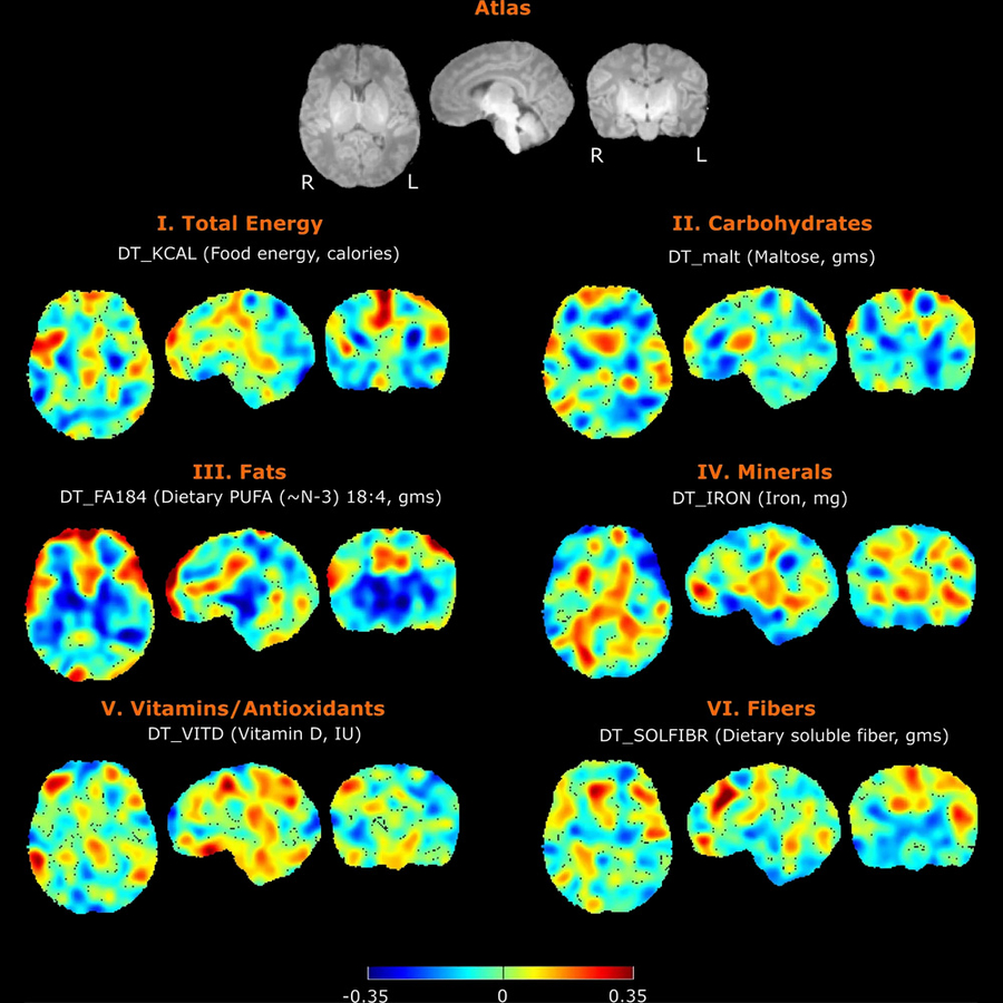
Nutrition has profound impact on neurodevelopment, but the mechanisms are only partly understood. In collaboration with other researchers at the Fetal-Neonatal Neuroimaging Developmental Science Center, we are studying the connections between maternal diet, breastmilk contents, infant brain development and child neurodevelopment. We also have a pilot study on the trajectory of infant develop the important skill of oral feeding with the goal of improving diagnosis and personalized care through quantitative EMG assessment of infant feeding, advanced computational analytics, and identifying biomarkers of neonatal outcomes.

Neurodevelopment FreeSurfer

Modifiers of Neurodevelopment among Patients with Congenital Heart Disease

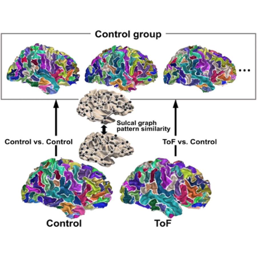
Congenital heart disease (CHD) is the most common severe malformation. As improvements in medical and surgical management have led to increased survival, patients with congenital heart disease face additional lifelong health risks. Neurodevelopmental delay or impairment is the most common extracardiac complication of CHD. To better understand the mechanisms of neurodevelopmental risk in patients with CHD, we have recently participated a clinical trial that collected genetic, clinical, and neuropsychological testing data. Ongoing projects include further analysis of that trial data, and local pilot studies.

Gene Discovery Data

Gene Discovery in Congenital Heart Disease

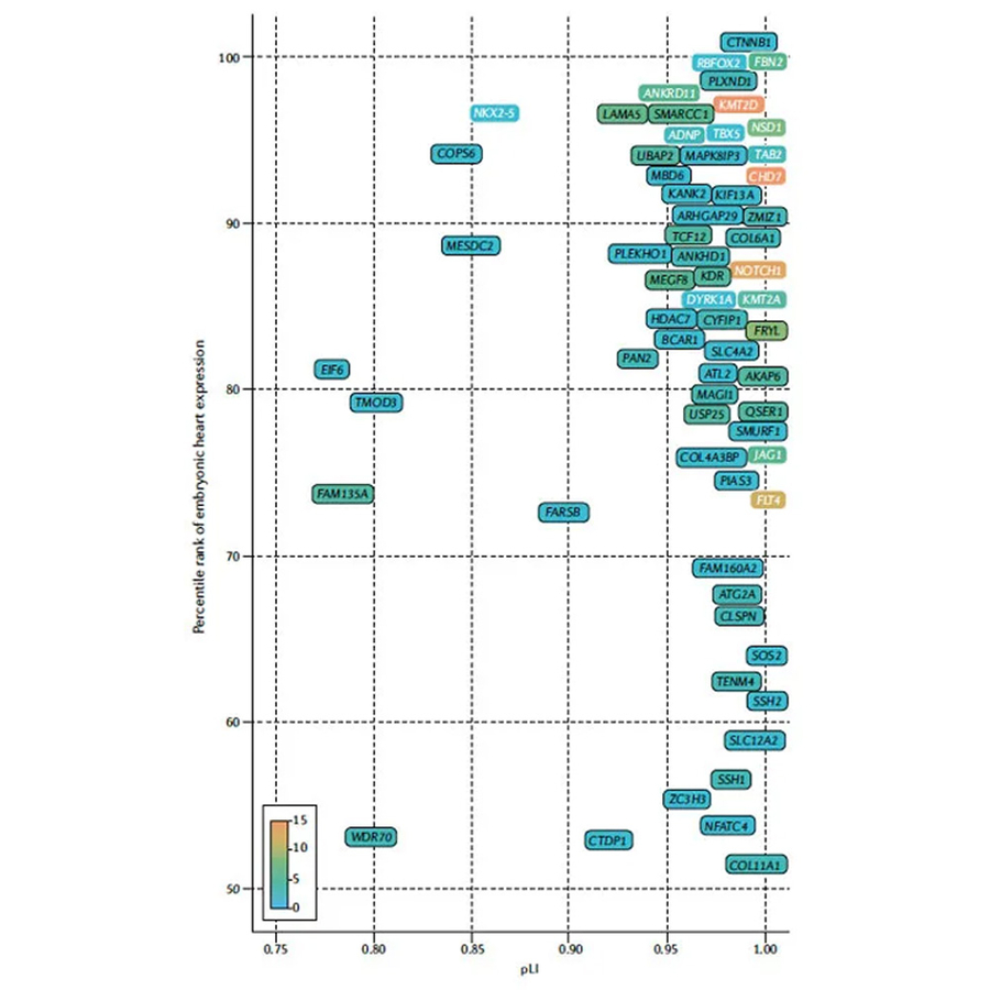
We study the genetics of congenital heart disease with the goal of improving diagnosis and personalized care through gene discovery, functional analysis of patient variants, and identifying biomarkers of neonatal outcomes. Approaches include computational biology projects, cell culture projects, and multi-omic analysis of patient samples.

Publications

  • Ladha, F. A., Avillach, P., Opotowsky, A. R., Nael, A., Brueckner, M., Chung, W. K., Cnota, J. F., Gelb, B. D., Lewis, M., Liu, C., Roberts, A. E., Seidman, C. E., Seidman, J. G., Tristani-Firouzi, M., Wagner, M., Newburger, J. W., Kim, Y., & Morton, S. U. (2026). Rare KDR Variants Define a Distinct Genetic Contribution to Congenital Heart Disease.. Circulation. Genomic and Precision Medicine, e005659. https://doi.org/10.1161/CIRCGEN.125.005659 (Original work published 2026)
  • Movassagh, M., Newbury, L., Hehnly, C., Whalen, A. J., Peterson, M., Mondragon-Estrada, E., Ericson, J. E., Smith, J., Sasanami, M., Natukwatsa, D., Mugamba, J., Ssenyonga, P., Onen, J., Burgoine, K., Zhang, L., Olupot-Olupot, P., Kumbakumba, E., Wegoye, E., Ochora, M., … Schiff, S. J. (2026). Genome admixture analysis of 1,030 Ugandan infants with neonatal sepsis and hydrocephalus demonstrates geographical stratification of population disease risk.. MedRxiv : The Preprint Server for Health Sciences. https://doi.org/10.64898/2026.03.16.26348489 (Original work published 2026)

    BACKGROUND: Neonatal disorders such as post-infectious hydrocephalus exhibit a higher incidence in Africa, where the intricate relationships between genetic ancestry, environmental exposures, and other risk factors likely contribute to the increased incidence.

    METHODS: To start to characterize the common genetic architecture of Ugandan infants, we analyzed genome sequencing data from 1,030 Ugandan infants recruited from studies targeting neonatal sepsis and hydrocephalus. We employed genetic admixture analysis and integrated geospatial data to examine the relationships between genetic backgrounds and disease prevalence within this cohort.

    RESULTS: Our results identified four distinct genetic admixture groups, each correlating strongly with specific geographic distributions across Uganda. Notably, a predominance of one admixture group, most common in northern Uganda, was overrepresented in the participants with post-infectious hydrocephalus.

    CONCLUSION: This study underscores the importance of genetic factors in disease manifestation at the population level, and a role for such precision public health approaches in complex neonatal disorders in African populations.

  • Lee, J., Wu, J., Pittman, M., Grant, Z. L., Kuang, S., Quiat, D., Morton, S. U., Fudenberg, G., Traglia, M., Hayes, K. A., Consortium, P. C. G., Kumar, R., Bruneau, B. G., & Pollard, K. S. (2026). Structural variants in human congenital heart disease disrupt distal genomic regulatory contacts of developmental genes.. BioRxiv : The Preprint Server for Biology. https://doi.org/10.64898/2026.02.28.708767 (Original work published 2026)

    Predicting the functional significance of structural variants (SVs) associated with genetic diseases remains challenging. To test the hypothesis that SVs from people with congenital heart disease (CHD) disrupt developmental chromatin interactions, we developed CardioAkita, a machine-learning model that predicts how variants alter 3D chromatin structure. Analyzing previously genotyped de novo SVs ( dn SVs), we observed a positive association between CHD severity and CardioAkita scores across dozens of families. From whole-genome sequencing of three individuals with CHD we predicted disruptive dn SVs. Induced pluripotent stem cells engineered to harbor these variants confirmed CardioAkita's predictions of 3D chromatin changes, and further revealed aberrant expression of local genes including cardiac developmental genes, suggesting that chromatin reorganization plays a significant mechanistic role in the genetic etiology of CHD. Our findings highlight the potential for models of 3D chromatin organization to predict the pathogenicity and underlying mechanisms of SVs in human disease.

  • O’Brien, E. A., Wypij, D., Rofeberg, V., Chamseddine, R., Velasco-Annis, C., Taylor, K., Pujols, K. H., Hart, N. J., Morton, S. U., Litwin, J., Kumar, N., Kennedy, K., Wilkins-Haug, L., Newburger, J. W., Odibo, A. O., Gholipour, A., Shimony, J. S., Rollins, C. K., & Ortinau, C. M. (2026). Brain volumes in fetuses with congenital heart disease and placental vascular abnormalities.. Journal of Perinatology : Official Journal of the California Perinatal Association. https://doi.org/10.1038/s41372-026-02601-4 (Original work published 2026)

    OBJECTIVE: Investigate the association between placental vascular abnormalities and regional brain volumes in congenital heart disease (CHD) fetuses with and without genetic abnormalities.

    STUDY DESIGN: Secondary analysis of brain magnetic resonance imaging (MRI) and placental pathology data from 121 CHD fetuses enrolled in prospective neuroimaging studies at two centers.

    RESULTS: Placental vascular abnormality was present in 46% of fetuses, and genetic abnormality was present in 19%, including 12% with both abnormalities. Fetuses with the combination of placental and genetic abnormalities had smaller brain volumes compared to fetuses without either abnormality for total brain, subcortical gray matter, brainstem, and cerebellum, with a significant interaction (P < 0.05) between placental and genetic abnormalities for intracranial and subcortical gray matter volumes.

    CONCLUSION: Smaller brain volumes for CHD fetuses with placental and genetic abnormalities may suggest common genetic pathways affect placental, heart, and brain development, or that genetic abnormalities heighten vulnerability when placental changes occur.

  • Ladha, F. A., VanderPluym, C., Mondragon-Estrada, E., Avillach, P., Brueckner, M., Chung, W. K., Cnota, J. F., Gelb, B. D., Lewis, M., Liu, C., Roberts, A. E., Seidman, C. E., Tristani-Firouzi, M., Wagner, M., Morton, S. U., Newburger, J. W., & Investigators, P. C. G. C. (2026). Prothrombin G20210A and Factor V Leiden Variants Are Not Associated With Thrombotic Events in Congenital Heart Disease: An Observational Trial.. Journal of the American Heart Association, e046959. https://doi.org/10.1161/JAHA.125.046959 (Original work published 2026)

    BACKGROUND: Thrombotic events, including acute ischemic stroke, are more common in individuals with congenital heart disease (CHD). Whether common thrombophilia variants contribute to thrombotic risk in this population remains unclear. We evaluated whether prothrombin G20210A (F2 c.97G>A) and factor V Leiden (F5 c.1601G>A; p.Arg534Gln) are associated with thrombotic events in CHD.

    METHODS: Participants in the Pediatric Cardiac Genomics Consortium with exome sequencing and electronic medical record data were identified. Individuals were stratified by prothrombin G20210A and factor V Leiden genotypes, ventricular physiology, and antithrombotic therapy. The primary outcome was the presence of International Classification of Diseases (ICD) or Phecodes (phenotype codes) for thrombotic events.

    RESULTS: Among 4008 participants (median age, 11.4 [interquartile range, 5.1-17.9] years; 44.4% boys), thrombotic events occurred in 737 (18%), including 93 (13%) with acute ischemic stroke. Compared with the Genome Aggregation Database, the CHD cohort had a lower prevalence of heterozygous prothrombin G20210A and factor V Leiden variants. Variant prevalence did not differ between participants with and without thrombotic events. Single-ventricle CHD was associated with higher thrombosis frequency than biventricular CHD (35% versus 16%, P≤0.0001), without differences in variant prevalence.

    CONCLUSIONS: In this multicenter CHD cohort, prothrombin G20210A and factor V Leiden were not significantly associated with thrombotic outcomes, supporting recommendations against routine screening. Given low variant prevalence, the study was powered to exclude only large associations. Reduced variant frequency suggests survivorship bias beginning in fetal life. Larger integrated clinical-genomic studies are needed to refine thrombotic risk stratification in CHD.

    REGISTRATION: URL: https://www.clinicaltrials.gov; Unique Identifier: NCT03347214.